Breaking news and analysis on politics, business, world national news, entertainment and more.
27+ Homer Wright Rosettes Usmle PNG
09/12/2018 00:00
27+ Homer Wright Rosettes Usmle PNG. Although the cellular mechanisms responsible for the formation of. Do not confuse with perivascular pseudorosettes/rosettes found in ependymomas.
Pathology Outlines Embryonal Rhabdomyosarcoma from www.pathologyoutlines.com
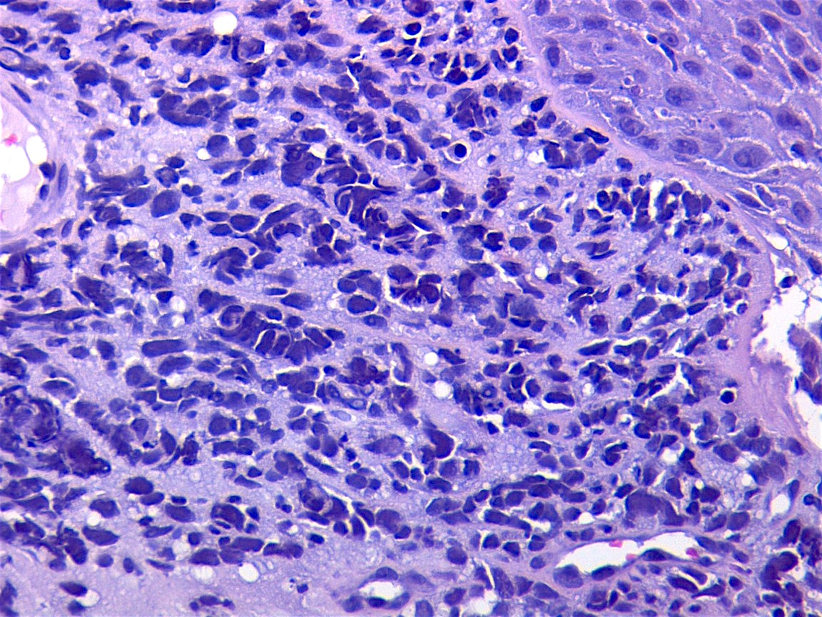
Two rosettes can be seen in this image along with foci of calcification. • presence of fibrous connective tissue stroma will produce a lobular pattern. Here the rosettes have a dense feltwork of interwoven cytoplasmic processes of nerve cells and neuroglial cells( neuropil) in the centre.
Homer wright rosettes are a type of rosette in which differentiated tumor cells surround the neuropil.
Homer wright rosette — a circular or spherical grouping of dark tumor cells around a pale, eosinophilic, central area that contains neurofibrils but lacks a lumen; Examples of tumors containing these are neuroblastoma, medulloblastoma, pinealoblastoma, and primitive neuroectodermal tumors of bone. This histologic finding is found in neuroblastoma, medulloblastoma and retinoblastoma. Although the cellular mechanisms responsible for the formation of.